

Founding Partner
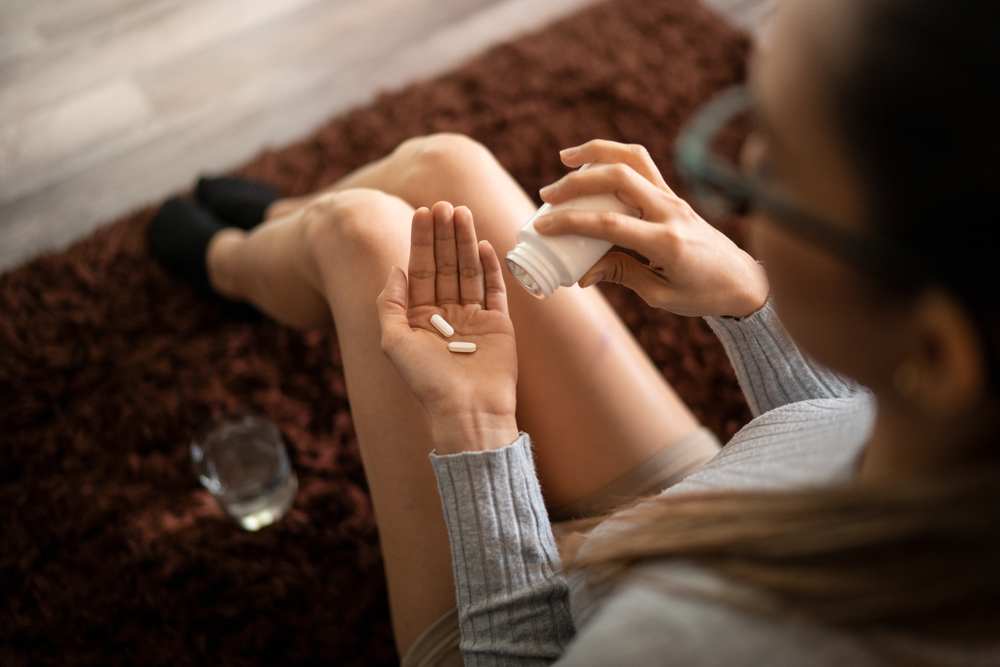
 Parker Waichman LLP is a national product liability law firm helping victims of injuries caused by defective and dangerous products. Our product liability lawyers tackle claims involving prescription drugs, defective medical devices, cosmetics, and any other potentially hazardous products in order to recover the compensation our clients need and deserve.
We’re not afraid to take on big companies, and we’ve built a reputation for getting results, to the tune of $2 billion and counting. If you have a potential defective product action, call our firm today for a free consultation with an experienced product liability attorney who can help you understand your legal rights.
Our skilled legal team has extensive experience litigating defective product cases, and we have successfully secured more than $2 billion in compensation for our clients. We help injured people receive product liability settlements and verdicts to cover their medical costs, make up for lost time at work, and compensate them for their pain and suffering. Over the course of decades handling these cases, our defective product law firm has received numerous honors from our colleagues, judges, and clients, including a listing in Best Lawyers and an AVVO rating of 9.8 out of 10.
When you have been harmed by a defective product, you need the best defective product injury attorney on your side, and when you work with a product liability lawyer with our firm, you can feel confident that we have the skill and knowledge to get the results you deserve.
Parker Waichman LLP actively litigates cases against the manufacturers of medical devices that have been found to be harmful to patients. Some of these include:
A hernia occurs when organs or tissue bulge through a weakened wall of tissue or muscle in the body. One example is a ventral hernia, in which a person’s intestines bulge through the abdominal wall. Hernias can usually only be repaired with surgery, and one of the surgical methods doctors use is implantation of polypropylene or polyester mesh. The mesh will roughly match the shape and size of the hernia, and it will have many tiny holes throughout it, like a window screen.
During the surgical procedure, the surgeon will make an incision near the site of the hernia, push the herniated tissue back in place, and reinforce the muscle wall using a piece of mesh. Scar tissue is then supposed to grow into the holes of the mesh, making a more rigid wall to prevent hernia recurrence. However, studies show that polypropylene and polyester mesh can cause serious complications when implanted in the human body. Hernia mesh has been known to erode, degrade, tear, perforate nearby organs, and cause severe infections. On top of that, its porous composition makes the mesh almost impossible to remove. Many patients require multiple additional surgeries, and some face lifelong side effects.
Parker Waichman LLP is pursuing product liability cases against multiple hernia mesh manufacturers, and we are currently taking new ones. If you had hernia repair surgery using synthetic mesh, contact our firm today for a free case consultation to see if you qualify for a lawsuit.
People who are prone to developing blood clots or deep vein thromboses (DVT) are often prescribed anticoagulants by their physicians. These drugs work to thin the blood to prevent clots, or thrombi, from building up and causing heart attacks, strokes or pulmonary embolism. But some patients do not respond favorably to anticoagulants or cannot take them because of other medical conditions.
In these situations, a doctor might recommend implantation of an inferior vena cava (IVC) filter. An IVC filter is a tiny metal implant that resembles the underside of an umbrella. It is implanted into a major vein in the body (the inferior vena cava) through a catheter inserted into the groin. Once the filter is placed, it is supposed to sit inside the vein and trap blood clots that travel up the leg to prevent them from going to other areas of the body, like the heart, lungs, and brain.
These filters are mostly designed to be temporary, but they are very difficult to retrieve. They can become lodged in the vein, puncture the vein, break apart, migrate out of position or perforate nearby organs or tissue. They can also become completely clogged with thrombi and cause serious medical complications. Studies suggest that nearly every IVC filter will fail, leaving patients at risk for significant injuries.
Parker Waichman LLP has offices nationwide, making it easy to access top-tier legal help no matter where you are. Our local teams are familiar with regional laws and courts, giving you a strategic advantage.
Ready to discuss your product liability case? Contact our nearest office today for a free consultation. Call now (516) 466-6500 or schedule an appointment online for a free consultation.
Our experienced medical product liability lawyers are investigating IVC filter cases and filing lawsuits against several manufacturers.If you had an IVC filter implanted, contact our firm today. We will review your case to see if you can file a product injury claim in court.
Parker Waichman LLP
 Amputation Injury
 Aviation Accidents
 Birth Injury
 Boat Accidents
 Burn Injury
 Catastrophic Injury Lawyer
 Clergy Sexual Abuse
 Construction Accidents
 DUI Injury
 Distracted Driving Accidents
 Hit-and-Run Accidents
 Lyft Accidents
 Nursing Home Abuse
 Pain and Suffering
 Rideshare Accidents
 Class Action Lawsuits
 Baby Injury Lawsuits
 Toxic Torts
 Defective Products
 Defective Medical Devices
 Defective Drugs
Sexual Abuse
Defective Drugs
Sexual Abuse  9-11 Victim Compensation
Traumatic Brain Injuries
9-11 Victim Compensation
Traumatic Brain Injuries Labor Law Injuries
 Scooter Accidents
 Spinal Cord Injuries
Workers' Compensation
Spinal Cord Injuries
Workers' Compensation Intentional Acts
Why Trust UsWith Your Case?
We Take Care of Everything
No Recovery = No Fees
Decades of Experience
Recovered Billions of Dollars
Respected by Our Peers
Amputation Injury
Aviation Accidents
Birth Injury
Boat Accidents
Burn Injury
Catastrophic Injury Lawyer
Clergy Sexual Abuse
Construction Accidents
DUI Injury
Distracted Driving Accidents
Hit-and-Run Accidents
Lyft Accidents
Nursing Home Abuse
Pain and Suffering
Rideshare Accidents
Class Action Lawsuits
Baby Injury Lawsuits
Toxic Torts
Defective Products
Defective Medical Devices
Defective Drugs
Sexual Abuse
9-11 Victim Compensation
Traumatic Brain Injuries
Labor Law Injuries
Scooter Accidents
Spinal Cord Injuries
Workers' Compensation
Intentional ActsIf you or a loved one has been injured in an accident or have been injured by another party in some other way, we are here to stand up for your rights. Our personal injury attorneys have been representing injury victims and their families in Long Island and throughout the nation since the early 1980s.
4.8 from 549 Reviews
Our law firm is ready to represent you in your injury case. We’ve helped many New York residents as well as those needing help nationwide. Contact our team for a free case consultation today.
I had a great experience with them , everyone was very helpful and sweet.
Michelle Murphy
4 months ago
Zarahi was very professional and very Quick and very knowledgeable i realy appreciated her patience and perseverance she Deserves 100 stars 🌟 but since i can only send 5 i Guess i will just have to send that truly yours Rashine Downs
Kush Three
6 months ago
VERY NICE WORK PLACE THEY HAVE BEEN GOOD TO MY MOM
Whitney Brinson
5 years ago
They treated me with tender love and care
Terrell Weaver
2 months ago
Wonderful people. They made the whole process if dealing with a gov’t agency so easy. Special compliments to Gina Viti
Michael Ross
2 months ago
I’m a 9/11 first responder, and I can honestly say that Parker Waichman made me feel like they had my best interest in my VCF case. Ms Candalino & Ms Viti are top notch in my book. I was constantly informed on the status of my case. I would definitely recommend Parker Waichman LLP to family and friends.
D D
6 years ago
We have the experience and the skilled litigators to win your case. Contact us and speak with a real attorney who can help you.
We handle mass torts cases nationwide. Please contact our office to learn more.